
CPD Handheld Diagnostic MSK Sonography/Ultrasound & Joint Manipulation
- David Lintonbon DO

- Apr 24, 2025
- 2 min read
Updated: May 26, 2025
12th - 14th September 2025, British College of Osteopathic Medicine, London NW3 5RR
Master the fusion of imaging and joint manipulation - This 3-Day course with Charles and David, will sharpen your diagnostic acumen & enhance your treatment precision.
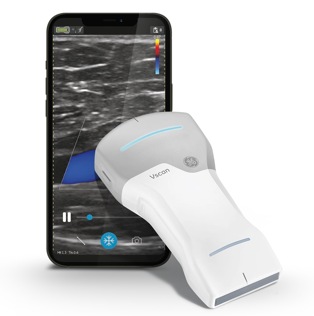
What is Vscan Air
Vscan Air is a compact, wireless ultrasound system that connects to tablets or smartphones— perfect for training in both classroom and real-world settings.
Key Benefits for Learners:
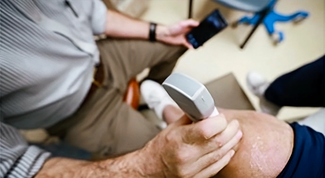
Real-Time, Hands-On Learning
Instant feedback during scanning—see anatomy and pathology live.
Practice scanning on peers, patients, or simulation models.
Portability & Flexibility
No bulky machines—train anywhere: classroom, clinic, sports field, or remote setting.
Encourages more scanning reps, which improves accuracy and confidence.
Enhanced Visualization
High-resolution imaging of musculoskeletal structures like tendons, ligaments, menisci, and joint effusions. Shows if joint manipulation is suitable or not.
Easy comparison with textbook anatomy or other diagnostic tools.
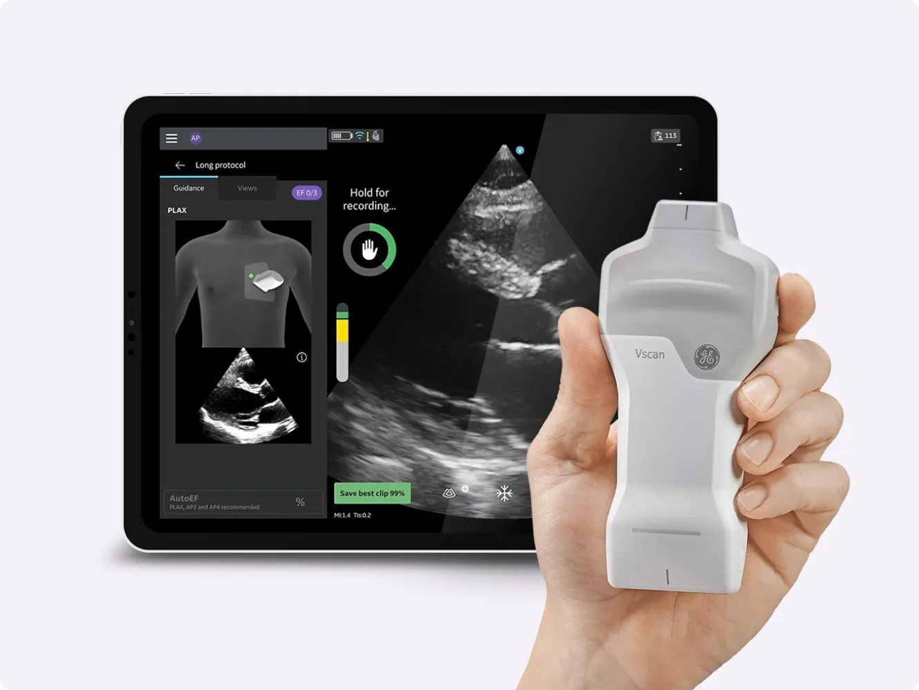
Built-In AI Tools & Cloud Sharing
Learners can store scans, annotate them, and get instructor feedback remotely.
Cloud-based case sharing supports collaborative learning.
Interdisciplinary Use
Ideal for physiotherapists, sports doctors, and emergency med students.
Encourages early adoption of point-of-care ultrasound (POCUS) skills.
Mini Case Study: Using Vscan Air for a Knee Injury Assessment
Setting: Sports Medicine Clinic | Instructor-Led Practical Session
Student: Samira, 3rd-year physiotherapy student
Patient: 22-year-old soccer player with acute right knee pain post-game
Initial Presentation:
Swelling and pain localised around the medial knee
No prior imaging available
Difficulty in full extension

Sonographic Assessment Using Vscan Air:
Step 1: Medial joint line scanned → Identified a hypoechoic fluid pocket consistent with joint effusion.
Step 2: MCL scanned in long axis → Found a partial fibre discontinuity, suggesting a Grade II MCL sprain.
Step 3: Meniscus visualised → No displacement or tear evident.
Step 4: Compared to contralateral knee for baseline anatomy.
Learning Outcome:
Samira correctly identified pathology and distinguished it from meniscal involvement.
Immediate feedback from instructor reinforced anatomical scanning planes.
Case saved to cloud and reviewed in class for group discussion on diagnosis and rehab plan.
Why This Matters:
The Vscan Air enabled real-time clinical correlation during a student-led assessment.
Students gained confidence in applying scanning protocols in acute settings.
Reinforced interdisciplinary learning: physiotherapy + ultrasound = faster, more informed treatment paths.

Comments